PET Scan for Cardiovascular Diseases: Diagnosis and Treatment
11 May, 2023
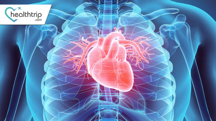
PET scans are commonly used for cancer diagnosis and staging, but they are also an important tool in diagnosing and treating cardiovascular disease. This blog reviews his use of PET scanning for cardiovascular disease, including its benefits, risks, and limitations.
What is cardiovascular disease?
Cardiovascular disease is a group of diseases that affect the heart and blood vessels, including coronary artery disease, heart failure, and arrhythmias. These conditions can cause a variety of symptoms such as chest pain, shortness of breath, and fatigue, and can lead to serious complications such as heart attacks and strokes.
Transform Your Beauty, Boost Your Confidence
Find the right cosmetic procedure for your needs.

We specialize in a wide range of cosmetic procedures

Diagnosis of cardiovascular disease
Diagnosing cardiovascular disease can be difficult because many of the symptoms are nonspecific and can be caused by other medical conditions. Additionally, traditional diagnostic tools such as electrocardiogram (ECG) and echocardiography do not always provide a clear picture of the underlying problem. This is where a PET scan can help. PET scans can provide detailed images of the heart and blood vessels, allowing doctors to detect abnormalities that other imaging tests cannot see. It can also be used to monitor treatment progress and assess the effectiveness of interventions.
How does PET scan work for cardiovascular disease?
Her PET scan for cardiovascular disease works similarly to his PET scan for cancer. A small amount of a radioactive substance called a radiotracer is injected into the patient's vein. The radiotracer travels to the heart and is taken up by the myocardium. A PET scanner detects positrons emitted from radioactive tracers and uses them to create a three-dimensional image of her heart.
Benefits of PET Scans in Cardiovascular Disease
One of the main advantages of PET scanning in cardiovascular disease is the ability to provide highly detailed images of the heart and blood vessels. This helps doctor’s spot problems that other imaging tests can't see. PET scans also help doctors determine the severity of a medical condition and determine the best course of treatment.
Another advantage of PET scanning is that it is non-invasive and does not expose you to radiation. This makes it a safer alternative to other imaging tests such as coronary angiography, which involves injecting a contrast agent into a blood vessel and exposing the patient to her x-rays.
Most popular procedures in India
Total Hip Replacemen
Upto 80% off
90% Rated
Satisfactory
Total Hip Replacemen
Upto 80% off
90% Rated
Satisfactory
Total Hip Replacemen
Upto 80% off
90% Rated
Satisfactory
ANGIOGRAM
Upto 80% off
90% Rated
Satisfactory
ASD Closure
Upto 80% off
90% Rated
Satisfactory

Risks and Limitations of PET Scans in Cardiovascular Disease
PET scans are generally safe, but they do come with some risks. Radiotracers used in PET scans emit small amounts of radiation and may increase the risk of cancer in some patients. However, radiation exposure from PET scans is relatively low, and the benefits of scanning generally outweigh the risks.
Another limitation of PET scans for cardiovascular disease is that they are not always covered by insurance. Some insurance plans consider PET scans to be experimental or exploratory and may not cover the cost of the test. Additionally, PET scans for cardiovascular disease can be expensive and are not available in all regions. Patients may need to go to a specialised centre for testing.
The Potential of PET Scans in Cardiovascular Disease
PET scans can be used to diagnose various cardiovascular diseases such as coronary artery disease, heart failure, and arrhythmias. Here are some ways to use PET scans for specific medical conditions.
1. Coronary artery disease
PET scans can be used to assess blood flow to the heart and detect blockages in coronary arteries. This helps doctors determine the best course of treatment, including: B. Angioplasty or bypass surgery.
2. Heart failure
PET scans can be used to assess heart function and determine the extent of damage caused by heart failure. This helps doctor’s plan treatment to improve heart function and reduce symptoms.
3. Arrhythmia
A PET scan can be used to identify the location and extent of abnormal electrical activity in the heart that is causing the arrhythmia. This helps doctors determine the best treatment options such as medications, implantable devices and ablation procedures.
4. Heart tumour
PET scans can be used to detect heart tumours and assess how far they have spread. This helps doctors decide the best course of treatment, such as surgery or radiation therapy.
5. Heart inflammation
PET scans can be used to detect heart inflammation caused by a variety of conditions, including myocarditis and sarcoidosis. This helps doctors determine the degree of inflammation and plan treatment to relieve symptoms and prevent complications. In addition to these specific conditions, PET scans can also be used to monitor treatment progress and assess the effectiveness of interventions. For example, PET scans can be used to determine whether a patient is responding to medication or to monitor the healing process after surgery.
Wellness Treatment
Give yourself the time to relax
Lowest Prices Guaranteed!

Lowest Prices Guaranteed!


